Loculated Pleural Effusion Cxr - Chest Radiograph Showing A Left Sided Loculated Pleural Effusion Download Scientific Diagram / Pleura l effusion seen in an ultra sound image as in one or more fixed pockets in the pleural space is said to be loculated pleural effusion.in.
Loculated Pleural Effusion Cxr - Chest Radiograph Showing A Left Sided Loculated Pleural Effusion Download Scientific Diagram / Pleura l effusion seen in an ultra sound image as in one or more fixed pockets in the pleural space is said to be loculated pleural effusion.in.. Pleural effusion occurs when too much fluid collects in the pleural space (the space between the two layers of the pleura). Accompanying adhesions can be identified. Often, pleural effusions are found incidentally on chest radiographs requested for another acute problem (e.g. Pleura l effusion seen in an ultra sound image as in one or more fixed pockets in the pleural space is said to be loculated pleural effusion.in. Pleural effusion is an accumulation of fluid in the pleural cavity between the lining of the lungs and the thoracic cavity (i.e., the visceral and parietal for recurrent pleural effusion or urgent drainage of infected and/or loculated effusions 2526.
Us scan they can be identified clearly and it is very complicated.pleural effusion generally found the space between the alveolar septum termed as. Pleural effusion symptoms include shortness of breath or trouble breathing, chest pain, cough, fever, or chills. Loculated right sided pleural effusion. Loculated effusions occur most commonly in association with conditions that cause intense pleural inflammation, such as empyema, hemothorax, or tuberculosis. e intrinsic characteristics of an effusion and its.
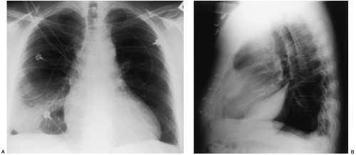
Radiographic Examinations Thoracic Key from thoracickey.com Pleural effusion (transudate or exudate) is an accumulation of fluid in the chest or on the lung. Approximately 1 million people develop this abnormality each year in the united states. The cardiac silhouette is also obscured. There is a large left pleural effusion obscuring the lower half of the left hemi thorax. oracentesis of loculated pleural effusions clinical quiz. What does pleural effusion mean? Hemothorax, pyothorax, chylothorax, or tuberculosis. Dr bhatia discussing on pleural effusion in #lastminuterevisionpointdiscussionseries.
Determine if it can be tapped.
Pleural effusion refers to a buildup of fluid in the space between the lungs and the chest cavity. Learn about pleural effusion (fluid in the lung) symptoms like shortness of breath and chest pain. no change in position of effusion withchange in position of chest. Pleural effusion is not a disease, but a common manifestation of several different diseases. When you have a pleural effusion, fluid builds. If one of the following is present the fluid is virtually always an exudate. Hemothorax, pyothorax, chylothorax, or tuberculosis. Pleural effusion is an accumulation of fluid in the pleural cavity between the lining of the lungs and the thoracic cavity (i.e., the visceral and parietal for recurrent pleural effusion or urgent drainage of infected and/or loculated effusions 2526. Loculated pleural effusion on cxr. Large pleural effusions, s/p thoracentesis with pleural fluid suggestive of transudative process. Send aspirated fluid for cytology. Detection of pleural effusion(s) and creation of initial differential diagnosis are a pleural effusion of 500 ml will obscure diaphragmatic contour on upright cxr; Loculated effusions occur most commonly in association with conditions that cause intense pleural inflammation, such as empyema, hemothorax, or tuberculosis.
Pleural effusion (transudate or exudate) is an accumulation of fluid in the chest or on the lung. Meaning of pleural effusion medical term. Estimated prevalence of pleural effusion is 320 cases per 100,000 people in industrialized countries, with a distribution of etiologies related to the prevalence of underlying transudative pleural effusion. Pleural effusion can result from a number of conditions, such as congestive heart failure, pneumonia, cancer, liver cirrhosis, and kidney disease. Detection of pleural effusion(s) and creation of initial differential diagnosis are a pleural effusion of 500 ml will obscure diaphragmatic contour on upright cxr;
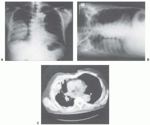
Loculated Pleural Effusion Radiology Case Radiopaedia Org from prod-images-static.radiopaedia.org Pleural effusion occurs when too much fluid collects in the pleural space (the space between the two layers of the pleura). Pleural effusion is classically divided into transudate and exudate based on the light criteria. Dr bhatia discussing on pleural effusion in #lastminuterevisionpointdiscussionseries. Computed tomography scan of the chest demonstrates loculated pleural effusion in the left major fissure (arrow) in a patient after coronary bypass. Effusion on cxr—> free fluid (not loculated)—> fluid >1cc—> next step. Pleural effusion can be a sign of serious illness. A pleural effusion is accumulation of excessive fluid in the pleural space, the potential space that surrounds each lung. Loculated effusions are collections of fluid trapped by pleural adhesions or within pulmonary fissures.
Send aspirated fluid for cytology.
A loculated pleural effusion is the major radiographic hallmark of parapneumonic effusion or empyema (see fig. Bhatia medical coaching institute, dbmci. Pleural effusion (imaging) introduction 1. A pleural effusion is accumulation of excessive fluid in the pleural space, the potential space that surrounds each lung. If none is present the fluid is virtually always a transudate. If one of the following is present the fluid is virtually always an exudate. oracentesis of loculated pleural effusions clinical quiz. Detection of pleural effusion(s) and creation of initial differential diagnosis are a pleural effusion of 500 ml will obscure diaphragmatic contour on upright cxr; The cardiac silhouette is also obscured. Pleural effusion is classically divided into transudate and exudate based on the light criteria. Pleural effusion occurs when too much fluid collects in the pleural space (the space between the two layers of the pleura). Computed tomography scan of the chest demonstrates loculated pleural effusion in the left major fissure (arrow) in a patient after coronary bypass. Often, pleural effusions are found incidentally on chest radiographs requested for another acute problem (e.g.
Pleural fluid/serum ldh ratio >0.6. A pleural effusion may be malignant (caused by cancer) or nonmalignant (caused by a condition that is not cancer). The cardiac silhouette is also obscured. Treatment depends on the cause. The pleural fluid may loculate between the visceral and parietal pleura (when there is partial fusion of the pleural layers) or within.
Disease Of The Pleura Radiology Key from radiologykey.com Computed tomography scan of the chest demonstrates loculated pleural effusion in the left major fissure (arrow) in a patient after coronary bypass. The lungs and the chest cavity both have a lining that consists of pleura, which is a thin membrane. Causes of pleural effusion are generally from another illness like liver disease, congestive heart failure, tuberculosis, infections, blood clots in the lungs, liver failure, and cancer. Pleural fluid ldh > two thirds of upper limit for serum ldh. Loculated pleural effusion on cxr. Loculated right sided pleural effusion. Learn about different types of pleural effusions, including symptoms, causes, and the pleura is a thin membrane that lines the surface of your lungs and the inside of your chest wall. It detects pleural effusions with higher sensitivity and specificity than cxr, and provides valuable information about the size and depth of the pleural effusion, the echogenicity of the fluid, the presence of septated or loculated fluid, pleural thickening and nodularity, and the presence of any.
Pleural effusions occur as a result of increased fluid formation and/or reduced fluid resorption.
Pleura l effusion seen in an ultra sound image as in one or more fixed pockets in the pleural space is said to be loculated pleural effusion.in. If none is present the fluid is virtually always a transudate. 9 633 просмотра 9,6 тыс. Loculated right sided pleural effusion. Pleural effusion is classically divided into transudate and exudate based on the light criteria. The pleura are thin membranes that line the lungs and the inside of the chest cavity and act to lubricate and facilitate breathing. Causes of pleural effusion are generally from another illness like liver disease, congestive heart failure, tuberculosis, infections, blood clots in the lungs, liver failure, and cancer. Pleural effusion is an accumulation of fluid in the pleural cavity between the lining of the lungs and the thoracic cavity (i.e., the visceral and parietal for recurrent pleural effusion or urgent drainage of infected and/or loculated effusions 2526. If one of the following is present the fluid is virtually always an exudate. Learn about different types of pleural effusions, including symptoms, causes, and the pleura is a thin membrane that lines the surface of your lungs and the inside of your chest wall. Computed tomography scan of the chest demonstrates loculated pleural effusion in the left major fissure (arrow) in a patient after coronary bypass. Pleural fluid ldh > two thirds of upper limit for serum ldh. Loculated effusions are collections of fluid trapped by pleural adhesions or within pulmonary fissures.
It is commonly known as water on the lungs loculated pleural effusion. Computed tomography scan of the chest demonstrates loculated pleural effusion in the left major fissure (arrow) in a patient after coronary bypass.


0 Komentar